

“My mammogram found calcifications. Should I worry?” In most cases, no.
Breast calcifications are tiny calcium deposits seen on a mammogram and are usually harmless. However, certain patterns may be associated with early breast cancer.
A breast cancer surgeon, together with the radiologist, determines whether additional imaging or a biopsy is needed based on the calcifications' appearance, not simply their presence.
What are breast calcifications?
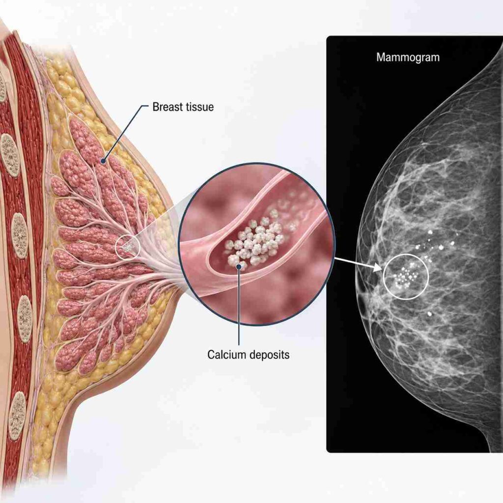
Breast calcifications are tiny calcium deposits that develop within breast tissue and appear as white spots on a mammogram. They usually cause no symptoms, cannot be felt during a breast examination and are most often benign. Their size, shape and distribution help determine whether further assessment is needed.
How do calcifications appear on a mammogram?
Breast calcifications on mammogram appear as small white dots or flecks because calcium blocks X-rays. They may be scattered, grouped or arranged in specific patterns. Radiologists evaluate their morphology and distribution to decide whether they represent a benign finding or require additional investigation.
Are breast calcifications caused by eating too much calcium?
No. Calcium in food, dairy products or supplements does not cause breast calcifications. These deposits form within breast tissue as a result of natural ageing, tissue repair or benign breast conditions, not because of excess calcium intake.
What causes calcifications in breast tissue?
Breast calcifications usually develop during normal ageing or healing within breast tissue. They are common mammographic findings and rarely indicate cancer. Understanding the causes of breast calcifications helps explain why most women require reassurance rather than treatment.
Age-related changes
As breast tissue ages, natural cell turnover and degeneration can leave behind tiny calcium deposits. These calcifications become more common after menopause and typically have a benign appearance that does not require treatment or biopsy.
Benign breast conditions
Several non-cancerous breast conditions can lead to calcifications. These include fibrocystic breast changes, fibroadenomas, breast cysts, duct ectasia and fat necrosis. Radiologists can often recognise these benign patterns based on their characteristic appearance.
Previous injury, surgery or breast treatment
Breast surgery, biopsy, radiation therapy or previous injury may leave calcium deposits as tissue heals. These healing-related calcifications are generally harmless and can usually be distinguished from suspicious changes by their appearance on mammography.
What is the difference between macrocalcifications and microcalcifications?
Macrocalcifications vs microcalcifications differ mainly in size and clinical significance. Macrocalcifications are larger deposits that are almost always benign, whereas microcalcifications are tiny specks that sometimes require closer evaluation depending on their pattern rather than their presence alone.
What are macrocalcifications?
Macrocalcifications are large, coarse calcium deposits commonly seen in women over 50. They usually result from normal ageing or benign breast changes and rarely require additional imaging or treatment.
Do breast calcifications mean cancer?
No. Most breast calcifications are benign (harmless) and do not mean you have cancer.
They are common, microscopic calcium deposits found on mammograms, often caused by ageing, past breast injuries, or normal cell turnover.
When can microcalcifications indicate early breast cancer?
Microcalcifications on mammogram may require further evaluation when they appear tightly grouped, irregular or arranged in a linear pattern.
These features can sometimes be associated with DCIS calcifications, although imaging alone cannot confirm cancer. A biopsy is needed for a definitive diagnosis.
Does finding calcifications mean the cancer has spread?
No. Breast calcifications do not indicate that cancer has spread.
They are imaging findings that may represent benign tissue changes or very early disease. If cancer is diagnosed, additional investigations determine its stage and whether it has spread beyond the breast.
Which breast calcification patterns may concern a radiologist?
Radiologists assess the overall pattern of calcifications rather than a single feature. Size, shape, distribution and changes over time help determine whether calcifications are likely to be benign or require biopsy.
Size and shape
Very fine, irregular or variable-shaped calcifications may require closer evaluation because they are more likely to be associated with abnormal cells. Larger, coarse and well-defined calcifications are usually benign.
Clustered calcifications
Clustered calcifications in breast tissue refer to multiple tiny deposits located within a small area. Although many clusters are benign, some patterns may require magnification views or biopsy to exclude early breast cancer.
Linear or branching calcifications
Linear or branching calcifications follow the course of the breast ducts. This distribution may be associated with abnormal ductal cell changes and often requires tissue sampling for accurate diagnosis.
Calcifications found only in one breast
New calcifications in one breast are assessed carefully because they represent a recent change. Radiologists compare them with previous mammograms and evaluate their pattern before recommending further investigations.
New or changing calcifications
Calcifications that increase in number or change in appearance over time deserve further assessment. Comparing current and previous mammograms helps determine whether the changes are stable, probably benign or require biopsy.
What do “benign,” “indeterminate” and “suspicious” calcifications mean?
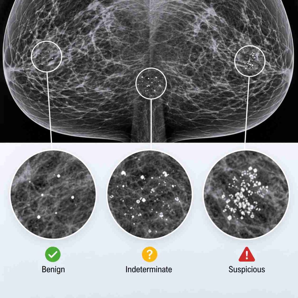
- Benign: Clearly non-cancerous calcifications that usually require no treatment.
- Suspicious: Irregular calcifications that may indicate early-stage breast cancer and need further evaluation.
- Indeterminate: Uncertain calcifications that require additional imaging or a biopsy to determine their cause.
Here’s a Detailed Breakdown
Benign-appearing calcifications
Benign calcifications have characteristic imaging features that are not associated with cancer. Can benign breast calcifications become cancerous? No. Benign calcifications themselves do not turn into cancer, although new unrelated calcifications can develop later.
Indeterminate calcifications
Indeterminate calcifications do not show clearly benign or suspicious features. Depending on their appearance, the radiologist may recommend a breast calcifications follow-up mammogram, additional diagnostic views or a biopsy for clarification.
Suspicious calcifications
Suspicious calcifications have imaging features associated with a higher likelihood of abnormal cells. Some may be reported as BI-RADS 4 calcifications, indicating that a biopsy is recommended because cancer cannot be ruled out based on imaging alone.
What does the BI-RADS score mean when calcifications are found?
The BI-RADS score is a standard reporting system used by radiologists to describe mammogram findings and recommend the next step. It helps ensure consistent communication about the likelihood of cancer and whether routine screening, follow-up imaging or biopsy is needed.
Why BI-RADS is included in the report
BI-RADS standardises mammogram reporting so every healthcare provider interprets the findings consistently. It also helps patients understand why no further testing, short-term follow-up or biopsy has been recommended.
BI-RADS categories patients commonly see
Patients with breast calcifications commonly encounter:
- BI-RADS 1: Normal mammogram.
- BI-RADS 2: Benign calcifications requiring routine screening.
- BI-RADS 3: Probably benign findings requiring short-term follow-up.
- BI-RADS 4: Suspicious findings where biopsy is recommended.
- BI-RADS 5: Findings highly suggestive of breast cancer.
What happens after calcifications are found on a screening mammogram?
Finding calcifications on a screening mammogram does not automatically mean cancer. The next step depends on how the calcifications appear. Many women simply need comparison with previous mammograms, while others require additional imaging or, less commonly, a biopsy.
Comparison with previous mammograms
Previous mammograms are reviewed to determine whether the calcifications are stable or newly developed. Calcifications that have remained unchanged over several years are more likely to be benign than those showing new or evolving patterns.
Diagnostic mammogram and magnification views
If additional assessment is needed, a diagnostic mammogram with magnification views provides a closer look at the calcifications. These specialised images help define their size, shape and distribution more clearly before deciding whether further testing is necessary.
Can ultrasound detect breast calcifications?
Ultrasound is generally not the best test for detecting breast calcifications because tiny calcium deposits are difficult to visualise. It may still be used to evaluate surrounding breast tissue if another abnormality is present.
When is a breast MRI considered?
Breast MRI is not routinely performed for calcifications alone. It may be recommended when additional information is needed about the extent of a known breast cancer or when a woman has a significantly increased risk of developing breast cancer.
When is a biopsy recommended for breast calcifications?
A breast biopsy is recommended when a mammogram shows suspicious microcalcifications that may indicate early breast cancer or precancerous changes. Radiologists assess their size, shape, pattern, and whether they have changed over time before recommending further testing.
Specific criteria for recommending a biopsy include:
- BI-RADS 4 or 5: Suspicious or highly suggestive of malignancy.
- Irregular appearance: Pleomorphic, linear, or branching calcifications.
- Clustered pattern: Tiny calcifications grouped closely together.
- New or changing calcifications: Compared with previous mammograms.
- Possible DCIS: Features associated with ductal carcinoma in situ or early-stage breast cancer.
When indicated, a stereotactic biopsy for breast calcifications is commonly performed because it accurately targets calcifications visible only on mammography.
If your results are suspicious, consult a breast cancer surgeon to discuss the diagnosis and appropriate next steps.
What can the biopsy result show?
A biopsy result provides a detailed microscopic examination of tissue to determine whether cells are normal, benign, precancerous, or cancerous. It helps confirm a diagnosis, assess disease severity, and guide the most appropriate treatment plan.
- Normal or benign tissue: Confirms healthy or benign breast tissue.
- High-risk or precancerous changes: Detects abnormal cells that may increase future cancer risk.
- DCIS: Identifies ductal carcinoma in situ (DCIS), an early-stage, non-invasive breast cancer.
- Invasive breast cancer: Confirms cancer has spread beyond the milk ducts and identifies its type.
- Infection or inflammation: Detects underlying infectious or inflammatory breast conditions.
What if the breast calcifications are benign?
Benign breast calcifications usually require no treatment because they are not cancerous.
Breast conservation surgery options are not needed for benign calcifications but may be discussed if a biopsy later confirms breast cancer.
If the calcifications are confirmed to be benign:
- Surgery is not required.
- Routine screening mammograms usually continue.
- Some women may need short-term imaging if stability has not yet been established.
- New breast symptoms should still be evaluated independently.
Questions to ask after calcifications appear on a mammogram
- Are these macrocalcifications or microcalcifications?
- Are they considered benign, indeterminate or suspicious?
- Are they new compared with my previous mammogram?
- What pattern or distribution did the radiologist see?
- What is my BI-RADS category?
- Do I need magnification views?
- Why is a biopsy being recommended?
- What type of biopsy will be performed?
- When should I receive the biopsy result?
- What follow-up will I need if the result is benign?
Breast calcifications need evaluation, not immediate panic
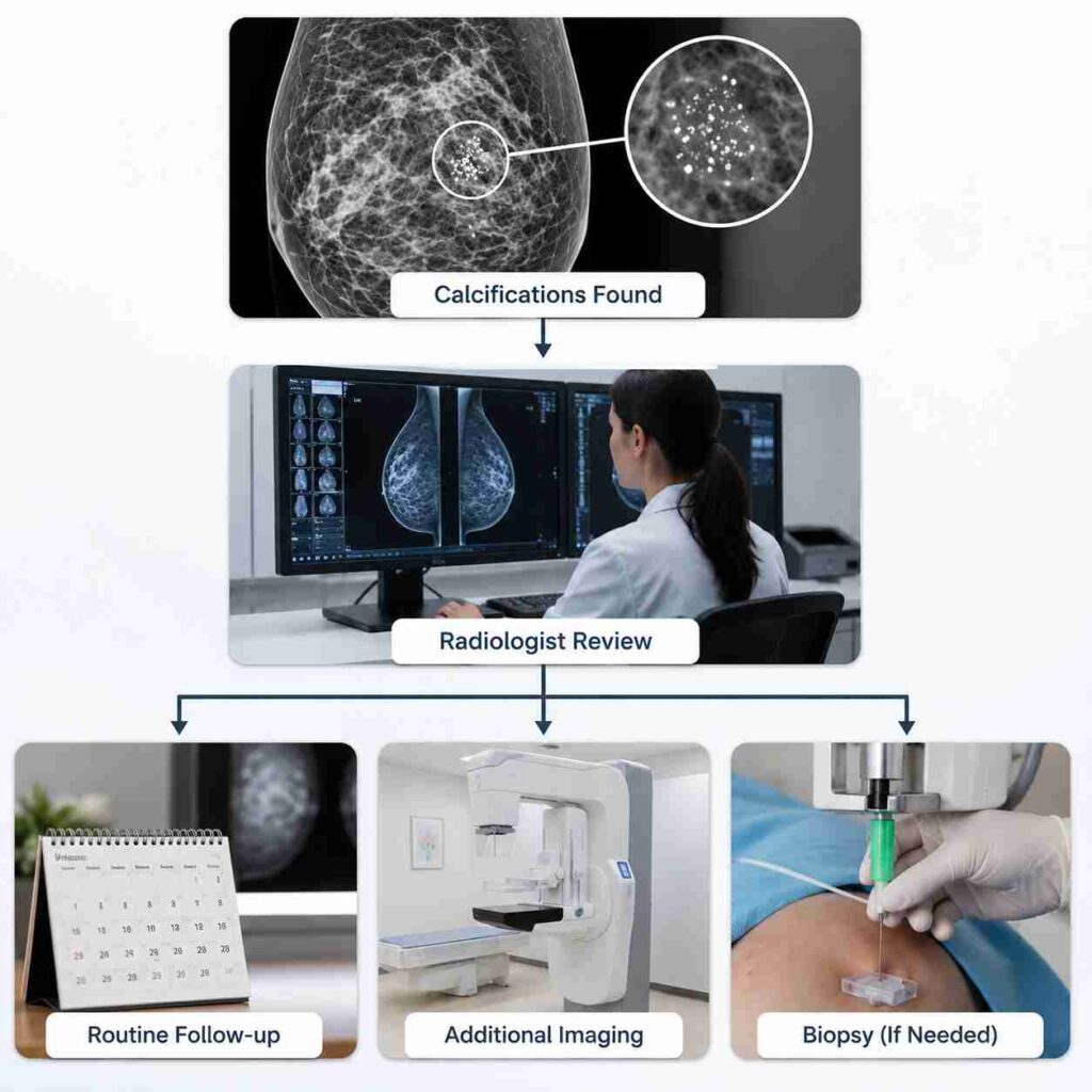
If your mammogram identifies calcifications requiring further evaluation, you can consult Dr Suvadip Chakrabarti, an experienced breast cancer surgeon in Kolkata, who can help you get an accurate diagnosis and timely, evidence-based treatment.
Call and book an appointment today.
People Also Ask
Should I worry about calcifications on my mammogram?
Not usually. Most breast calcifications are benign and require no treatment. Your radiologist will recommend additional imaging or biopsy only if the calcification pattern appears suspicious.
What percentage of breast calcifications are cancerous?
Only a small proportion are cancerous. What percentage of breast calcifications are cancer depends on their imaging characteristics, but the majority seen on screening mammograms are benign.
Are clustered breast calcifications always cancer?
No. Many clusters are benign. Their size, shape, distribution and stability determine whether further imaging or biopsy is required.
Can microcalcifications be benign?
Yes. Most microcalcifications are benign. Only certain patterns, such as irregular clustered deposits, require additional investigation.
What does BI-RADS 4 calcification mean?
BI-RADS 4 indicates that the calcifications are suspicious enough to recommend biopsy. It does not confirm cancer but means cancer cannot be excluded based on imaging alone.
Can breast calcifications disappear?
Most breast calcifications remain stable over time. They usually do not disappear, but stable benign calcifications generally require no treatment.
Do breast calcifications cause pain or a lump?
No. Breast calcifications almost never cause pain or a palpable lump. They are usually detected only during mammography.
Does a biopsy for calcifications mean I have cancer?
No. A biopsy is performed to determine the exact cause of the calcifications. Many biopsies confirm benign breast changes rather than cancer.